
Brachial Plexus: Find the Lesion
An interactive brachial plexus from roots to terminal nerves: place a lesion anywhere, see which muscles and which skin are lost on the patient beside it, and solve twelve clinical cases.
What you can do
- Explore the plexus from the C5 to T1 roots through trunks, divisions and cords to the terminal nerves.
- Place a lesion anywhere, preganglionic or postganglionic, alone or several at once.
- See the lost muscles and the sensory loss on the patient, palm and dorsum.
- Load classic patterns: Erb, Erb with root avulsion, extended Erb, Klumpke, cord lesions and the whole plexus.
- Solve twelve clinical cases in Find mode.
- Review neonatal brachial plexus palsy, the Narakas groups and the decisions.
Key concepts
Root avulsions (preganglionic) also take out the branches that leave the roots, such as the dorsal scapular and long thoracic nerves; a postganglionic lesion spares them.
Erb palsy is an upper trunk (C5-C6) lesion; Klumpke palsy involves C8-T1 and the hand.
The pattern of lost muscles and skin localizes the lesion to a root, trunk, cord or terminal nerve.
Teaching notes
The brachial plexus transmits all motor and sensory traffic between the cervical spinal cord and the upper limb. Because its fibers are reorganized into trunks, divisions, cords, and terminal nerves as they travel from neck to axilla, the pattern of muscle weakness and sensory loss allows precise localization of a lesion anywhere along that path.
Anatomy of the Plexus
The plexus is formed by the ventral rami of C5 through T1. These five roots unite into three trunks (upper, middle, lower), each trunk splits into anterior and posterior divisions, and the divisions regroup into three cords (lateral, posterior, medial) named by their relationship to the axillary artery. The cords give rise to the five major terminal nerves: musculocutaneous, axillary, radial, median, and ulnar.
Short branches leave the plexus before the trunks and cords are formed. The dorsal scapular nerve (rhomboids) and the long thoracic nerve (serratus anterior) arise directly from the C5 root and the C5-C7 roots respectively. This proximal origin is the key to distinguishing preganglionic from postganglionic injury.
Preganglionic versus Postganglionic Injury
A preganglionic injury, or root avulsion, tears the root from the spinal cord before those early branches have left. As a result, serratus anterior is paralyzed (scapular winging on wall push), the rhomboids are weak, the phrenic nerve may be involved (elevated hemidiaphragm on radiograph), and the cervical sympathetic chain at T1 may be disrupted (ipsilateral Horner syndrome: ptosis, miosis, enophthalmos). Because the motor neuron cell body is destroyed, spontaneous recovery is impossible and direct repair is not feasible.
A postganglionic injury leaves those root branches intact. Serratus and the rhomboids remain strong, the diaphragm is normal, and there is no Horner syndrome unless the lower cord is also involved. Postganglionic ruptures are amenable to nerve grafting, and neuropraxic lesions may recover spontaneously.
- Winging on wall push: long thoracic palsy, avulsion or very proximal lesion
- Ipsilateral Horner (ptosis, miosis): lower-root preganglionic injury at T1
- Elevated hemidiaphragm: phrenic involvement, associated with C5 avulsion
- Apical extrapleural hematoma on chest radiograph: strongly suggests C8-T1 avulsion
- Severe burning pain: hallmark of preganglionic injury; no potential for spontaneous recovery
Classic Injury Patterns
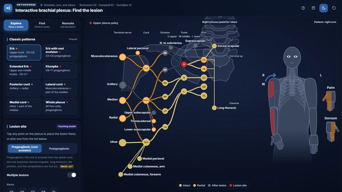
Upper plexus palsy (Erb-Duchenne) involves C5 and C6. The shoulder will not elevate or externally rotate, and elbow flexion is lost; the hand is largely intact. The typical posture is adduction, internal rotation, and an extended elbow. When C7 is also injured (extended upper palsy), elbow extension and wrist extension are additionally weak, but the lower trunk remains intact so grip and finger intrinsics are preserved.
Lower plexus palsy (Klumpke) involves C8 and T1. The hand is weak and clawed, with loss of intrinsic function and finger flexion, while the proximal shoulder-girdle muscles are spared. When the lower roots are avulsed, Horner syndrome is present.
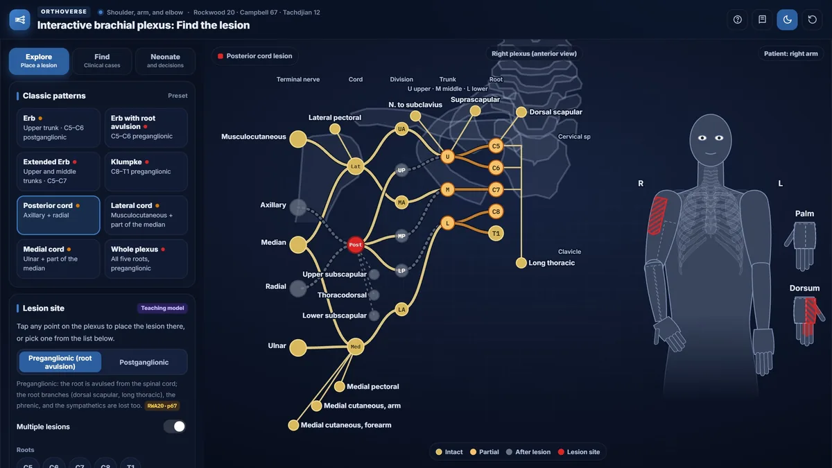
Cord lesions produce characteristic patterns. The posterior cord gives rise to the axillary and radial nerves, so a posterior cord lesion eliminates deltoid, triceps, and all extensors while sparing biceps and the finger flexors. The lateral cord supplies musculocutaneous fibers and the lateral root of the median nerve; its injury weakens elbow flexion, pronation, and wrist flexion, with loss of lateral forearm sensation. The medial cord carries the ulnar nerve and the medial root of the median nerve; its lesion produces intrinsic paralysis and a claw hand with the extensors intact. Cord lesions spare the muscles whose nerves leave before the cords form: serratus anterior (long thoracic), the rhomboids (dorsal scapular) and supraspinatus and infraspinatus (suprascapular, from the upper trunk). Pectoralis major and latissimus dorsi may be weak or spared, depending on whether the lesion lies proximal or distal to the pectoral and thoracodorsal branches.
Localizing the Lesion
Systematic examination of muscle groups and sensory territories, combined with clinical signs such as winging, Horner syndrome, and diaphragm elevation, allows accurate localization. The simulator displays which muscles are paralyzed or weakened and which sensory areas are lost for any point on the plexus, providing immediate feedback between anatomy and clinical findings.
Key movements to assess in sequence are shoulder abduction and external rotation, elbow flexion and extension, wrist extension, finger extension, grip strength, and hand intrinsics. Reflexes (biceps, brachioradialis, triceps) are abolished by complete nerve division but are unreliable in partial lesions.
Neonatal Brachial Plexus Palsy
Birth palsy follows traction on the plexus during delivery. In vertex delivery the upper plexus is stretched by lateral traction on the neck; in breech delivery the lower plexus is at risk. The Narakas classification describes four anatomic groups: group 1 (C5-C6), group 2 (C5-C7), group 3 (pan-plexus), and group 4 (pan-plexus with Horner syndrome, indicating T1 sympathetic injury). In one series, infants with no elbow flexion at three months all eventually regained some flexion, but the later the recovery, the worse the result.
Monitoring passive external rotation of the shoulder is critical in early infancy; a sudden decrease in passive external rotation signals posterior subluxation and should prompt ultrasound or imaging. If plexus surgery is being considered, the window for most procedures is between five and twelve months of age. For the external rotators, microsurgical reconstruction of the suprascapular nerve has not been reliable, and tendon transfer is usually required.
Using the Simulator
The Explore mode allows placement of a single lesion or multiple lesions anywhere on the plexus, with immediate display of paralyzed and weakened muscles and the affected sensory areas. Classic patterns can be loaded from the Preset list to compare expected findings with the anatomy. The Find mode presents twelve clinical cases: the learner reads the history and examination, then taps the lesion site on the plexus diagram. An exact answer scores two points; a near answer (correct level but not exact) scores one point. The Neonate mode displays the Narakas groups, mechanism, natural history, and decision points from the textbook.
Try this in the simulator
- In Explore mode, tap a C5-C6 root lesion, then switch between Preganglionic and Postganglionic using the Root lesion level toggle, and compare which muscles change.
- Load the Classic patterns Posterior cord lesion and observe that the deltoid and triceps are both lost while biceps and grip remain intact.
- Open the Movements panel and tap Shoulder abduction with a C5 root lesion placed; note that abduction is not completely lost because supraspinatus remains functional.
- In Find mode, work through all twelve clinical cases and use the Score display to track your exact and near answers.
- Switch to Neonate and decisions, select Group 4, and read the decision points for a pan-plexus injury with Horner syndrome.
Pitfalls
- Full shoulder abduction is still possible with a paralyzed deltoid because supraspinatus and scapular rotation compensate; always palpate the deltoid during testing.
- Brachioradialis can maintain elbow flexion even when biceps is completely paralyzed; palpate the biceps belly during the flexion test to avoid missing the diagnosis.
- A lower-trunk lesion alone does not produce Horner syndrome; ipsilateral Horner requires preganglionic injury at the T1 root.
- In a cord lesion, serratus anterior, the rhomboids, supraspinatus and infraspinatus are spared; weakness of these muscles points to a root or upper-trunk injury.
- Tendon reflexes are unreliable indicators in partial plexus lesions; complete nerve division abolishes the reflex, but partial injury may leave it present.
One-page high-yield card
The key facts, numbers and pitfalls of this simulator on one printable page. Free for personal study.
Further reading
- Rockwood and Green's Fractures in Adults, 10th edition: ch 20 (nerve injuries and the brachial plexus).
- Campbell's Operative Orthopaedics, 15th edition (2026): ch 67 (peripheral nerve injuries).
- Tachdjian's Pediatric Orthopaedics: ch 12 (brachial plexus birth palsy).
Designed for Orthoverse by Dr. M. M. Bakhshaei. Built to make orthopedic concepts visible, measurable and easy to explore.
Have an idea for the next simulator?
Tell us which concept you would like to explore. Message our bot and we will read every idea.
For medical education only. Orthoverse simulators are teaching models for health professionals and students. They are simplified on purpose, are not medical devices and are not a substitute for clinical judgement, patient-specific imaging or the original literature.
© 2026 Orthoverse. All rights reserved. The simulators, their code, drawings and models are original work. Free to use for learning on orthoverse.org; copying, embedding, scraping or redistribution is not permitted. Textbook references are cited for further reading only.