
Compartment Syndrome and Fasciotomy
An interactive cross section of the leg, thigh, forearm and foot: learn every compartment, calculate delta P from compartment and diastolic pressure, and plan the fasciotomy incisions that release them.
What you can do
- Tap any compartment of the leg to see its muscles, nerves, vessels and boundaries.
- Switch to the thigh, forearm and foot cross sections.
- Calculate delta P from compartment pressure and diastolic blood pressure, with time and trend.
- Load eleven cases, from borderline delta P to the child with a higher baseline pressure.
- Plan the standard two-incision leg fasciotomy and see what a single lateral incision misses.
- Check yourself with a quiz on pressures, compartments and incisions.
Key concepts
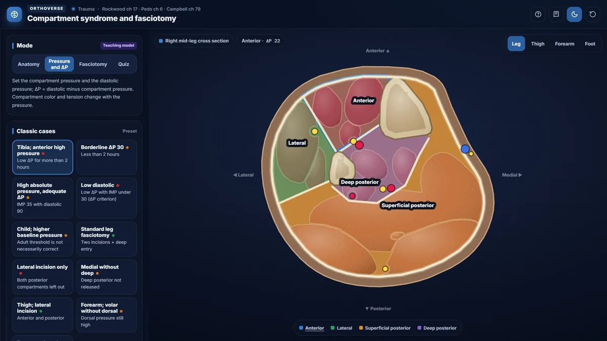
The leg has four compartments: anterior, lateral, superficial posterior and deep posterior; each has its own contents and boundaries.
Delta P, diastolic pressure minus compartment pressure, reflects perfusion better than an absolute compartment pressure; a clear clinical picture needs no measurement.
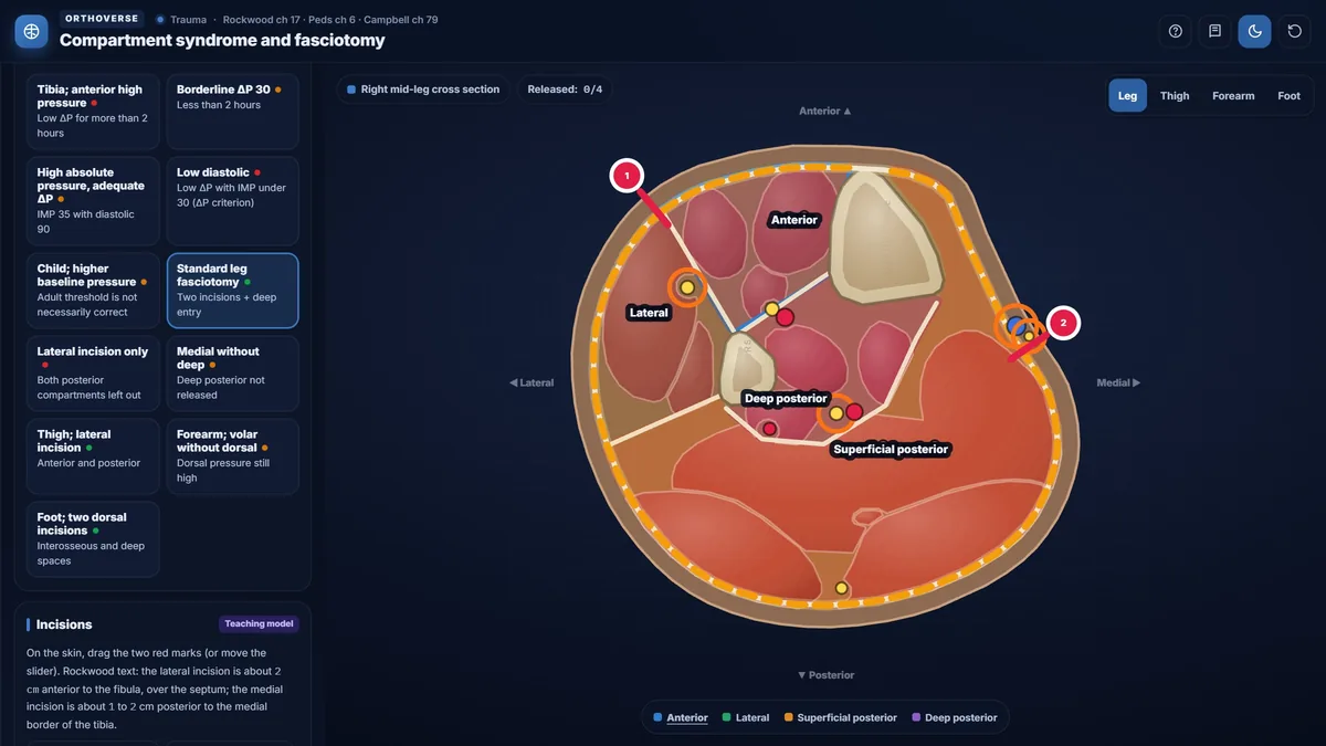
The standard leg fasciotomy uses two incisions, anterolateral and posteromedial, and the deep posterior compartment must be opened deliberately.
Teaching notes
Acute compartment syndrome (ACS) is a surgical emergency in which rising pressure within a closed fascial space impairs perfusion of the contained muscles and nerves. Irreversible muscle loss begins within hours of ischemia onset, making prompt recognition and complete fasciotomy decisive.
Compartmental anatomy
The leg has four compartments: anterior, lateral, superficial posterior, and deep posterior. Each is a distinct fascial envelope with its own muscle groups, nerves, and vessels. The deep posterior compartment contains tibialis posterior, flexor digitorum longus, flexor hallucis longus, and the posterior tibial nerve and vessels; it must be opened deliberately during fasciotomy because it is not reached by a lateral skin incision alone.
The thigh has anterior, posterior, and adductor compartments. The forearm has volar (superficial and deep layers), mobile wad, and dorsal compartments. The foot has interosseous spaces, medial, central, and adductor hallucis compartments, with the calcaneal compartment at hindfoot level.
- Anterior leg: tibialis anterior, EDL, EHL, deep peroneal nerve, anterior tibial vessels.
- Superficial posterior leg: gastrocnemius, soleus, plantaris, sural nerve.
- Deep posterior leg: tibialis posterior, FDL, FHL, posterior tibial nerve and vessels.
Clinical diagnosis
Pain out of proportion to the injury and pain with passive stretch of compartment muscles are the earliest clinical findings. Paresthesia, weakness, swelling, and loss of pulse are progressively later. Paralysis and absent pulse are late signs that carry a poor prognosis; they must never be awaited as a threshold for intervention.
In young children the classical signs are unreliable. The three A's — anxiety, agitation, and an increased analgesic requirement — are the clinical alerts. Normal resting compartment pressure is higher in children (13.3 to 16.6 mm Hg) than in adults (5.2 to 9.7 mm Hg), so the adult absolute-pressure threshold does not transfer directly to a child.
- One positive sign: probability of confirmed ACS is less than 26%.
- Three or more findings: probability exceeds 90%, but paralysis is already present.
Pressure measurement and the delta P criterion
Delta P equals diastolic blood pressure minus compartment pressure. The Rockwood threshold for adults is a delta P at or below 30 mm Hg. A single measurement is insufficient in most patients; the pressure trend and the duration of low delta P matter. The Edinburgh protocol calls for immediate fasciotomy when delta P remains at or below 30 mm Hg for more than two hours with a rising compartment pressure.
When the clinical picture is unambiguous, Campbell states that pressure measurement is not required and the surgeon should open the compartment without delay. When the patient is not alert and examination is unreliable, serial pressure monitoring becomes essential.
- Muscle dysfunctional after 2 to 4 hours of ischemia; irreversible loss after 4 to 12 hours.
- Nerve injury: abnormal after 30 minutes; irreversible after 12 to 24 hours.
Leg fasciotomy technique
The standard two-incision technique releases all four leg compartments. A lateral incision approximately 2 cm anterior to the fibula, over the septum between the anterior and lateral compartments, gives direct access to both fasciae. A medial incision approximately 1 to 2 cm posterior to the medial tibial border allows release of both posterior compartments; the surgeon must retract the superficial compartment posteriorly and elevate it off the tibia to enter the deep compartment.
The superficial peroneal nerve exits the fascia approximately 10 cm above the lateral malleolus and must be identified during the lateral incision. In the medial incision field, the saphenous vein and nerve require identification and protection, as do the posterior tibial vessels and nerve.
Fasciotomy in other regions and wound care
For the thigh, a single lateral incision releases anterior and posterior compartments; a separate medial incision is added when the adductor compartment requires decompression. For the forearm, a volar incision from the lacertus fibrosus to the carpal tunnel, with the deep interval opened between flexor carpi ulnaris and flexor digitorum superficialis, is performed first; if dorsal pressure remains elevated, a straight dorsal incision follows. For the foot, two dorsal incisions over the second and fourth metatarsals decompress the interosseous spaces; a medial incision addresses the calcaneal compartment in hindfoot injuries.
Fasciotomy wounds must never be closed primarily; compartment pressure may remain elevated. A second-look at approximately 48 hours allows closure only when all muscle groups are confirmed viable.
Try this in the simulator
- In Anatomy mode, tap the deep posterior compartment on the leg cross-section to review its contents and stretch signs before switching to Fasciotomy mode.
- In Pressure and delta P mode, load the 'Low delta P for more than 2 hours' preset and observe how compartment color and the pressure bar change; note the Live result panel.
- In Fasciotomy mode, place a single lateral incision only and tap 'Perform fasciotomy' to see which compartments remain unreleased, then add the medial incision to complete all four releases.
- Switch to the thigh, forearm, and foot cross sections using the Select compartment control and compare how incision strategies differ by region.
- Use Quiz mode and, for anatomy questions, tap directly on the cross-section diagram to identify compartments by location.
Pitfalls
- A palpable distal pulse does not exclude ACS; arterial flow can persist while compartment perfusion is critically impaired.
- A single lateral leg incision leaves both posterior compartments unreleased; all four must be decompressed.
- A short or subcutaneous fasciotomy incision fails to expose all muscle bellies and leaves the skin as a constricting band.
- Do not apply the adult delta P threshold to a child without integrating the full clinical picture and recognizing the higher normal resting pressures.
- Primary closure of fasciotomy wounds is contraindicated; always leave open and perform a second-look at approximately 48 hours.
One-page high-yield card
The key facts, numbers and pitfalls of this simulator on one printable page. Free for personal study.
Further reading
- Rockwood and Green's Fractures in Adults, 10th edition: ch 17 (compartment syndrome).
- Rockwood and Wilkins' Fractures in Children: ch 6 (compartment syndrome in children).
- Campbell's Operative Orthopaedics, 15th edition (2026): ch 79 (compartment syndrome and fasciotomy).

Designed for Orthoverse by Dr. M. M. Bakhshaei. Built to make orthopedic concepts visible, measurable and easy to explore.
Have an idea for the next simulator?
Tell us which concept you would like to explore. Message our bot and we will read every idea.
For medical education only. Orthoverse simulators are teaching models for health professionals and students. They are simplified on purpose, are not medical devices and are not a substitute for clinical judgement, patient-specific imaging or the original literature.
© 2026 Orthoverse. All rights reserved. The simulators, their code, drawings and models are original work. Free to use for learning on orthoverse.org; copying, embedding, scraping or redistribution is not permitted. Textbook references are cited for further reading only.