
3D Meniscus Atlas
A rotating 3D model of the tibial plateau and both menisci: tear patterns, vascular zones, repair techniques and meniscectomy, all on the same model.
What you can do
- Rotate and zoom a 3D tibial plateau with the medial and lateral menisci, their roots, the ligaments and the capsule.
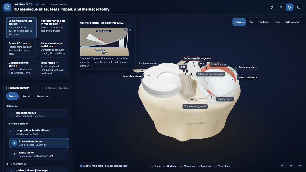
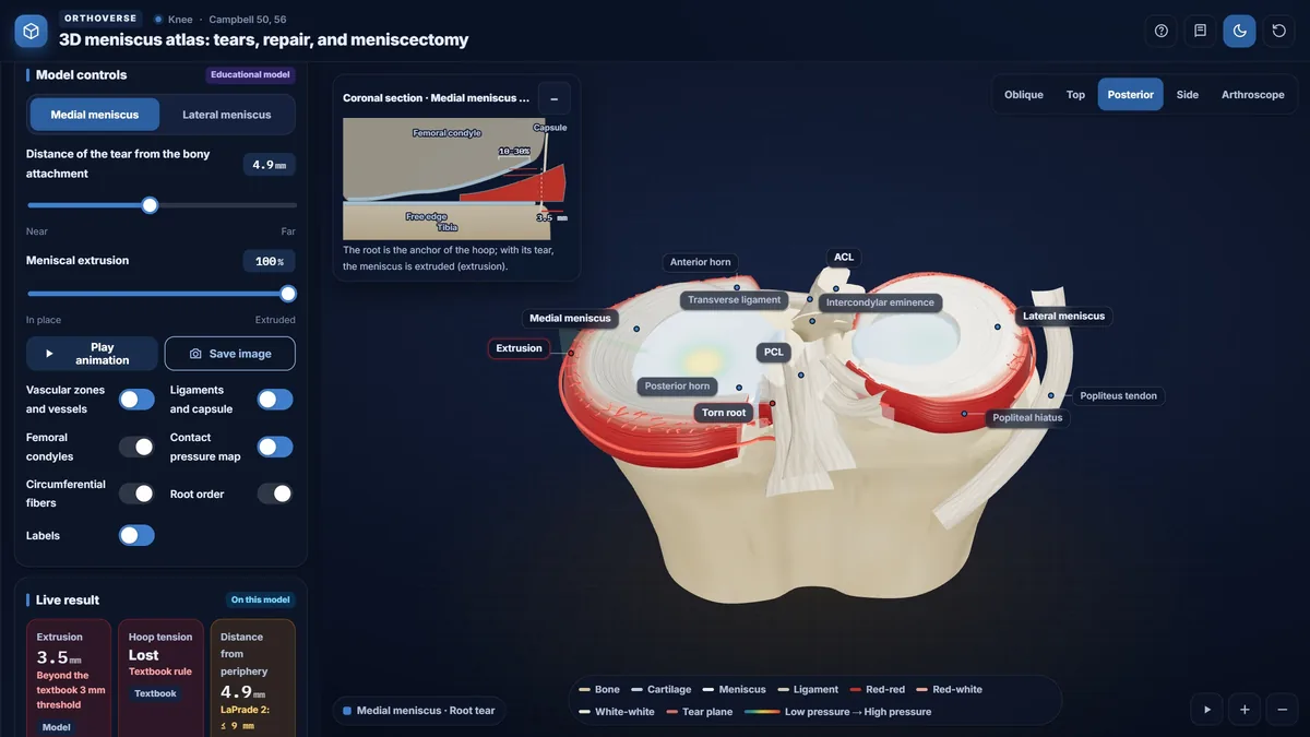
- Open six classic cases: bucket-handle locking, posterior root tear with extrusion, ramp lesion, lateral radial tear, parameniscal cyst and an ideal repair.
- Browse the tear library: longitudinal, bucket-handle, ramp, horizontal, oblique, radial, flap, complex and root tears.
- Show the vascular zones, the circumferential (hoop) fibers and a contact-pressure map.
- Step through repairs (inside-out, outside-in, all-inside, transtibial root repair) and resections (partial, subtotal, total).
- Switch views: oblique, top, posterior, side and an arthroscopic view from the anterior portal.
- Read live values for extrusion, hoop continuity and tear length, with the textbook numbers beside them.
Key concepts
The menisci are wedge-shaped and cover one-half to two-thirds of the articular surface. Their circumferential fibers turn axial load into hoop tension; a radial tear that reaches the capsular margin, or a root tear, breaks that ring.
Vessels reach only the outer 10% to 30% of the medial and 10% to 25% of the lateral meniscus, which is why peripheral tears in the red zone heal best.
Contact stress rises with the amount of meniscus removed: after medial meniscectomy the contact area falls by 50% to 70%, and lateral meniscectomy raises contact stress by 200% to 300%.
Teaching notes
The menisci are fibrocartilaginous wedges covering one-half to two-thirds of the tibial articular surface, transmitting 40% to 60% of load during standing. Their circumferential collagen fibers convert axial compression into hoop tension directed to the tibial roots; any disruption of that ring has immediate consequences for contact stress.
Anatomy and Biomechanics
Each meniscus has a triangular cross-section: the inferior surface is flat while the superior surface is concave, conforming to the femoral condyle. The thick peripheral border attaches via the coronary ligament; the inner border is free. The medial meniscus is C-shaped, firmly attached to the deep MCL throughout its periphery, and less mobile. The lateral meniscus is nearly circular, more mobile, and lacks capsular attachment at the popliteal hiatus. Collagen fibers run predominantly circumferentially; loading perpendicular to this orientation reduces tensile strength to less than 10% of the circumferential value.
- Lateral meniscus bears approximately 70% of lateral compartment load; medial bears roughly half of medial compartment load.
- Perimeniscal vessels supply only 10% to 30% of medial and 10% to 25% of lateral meniscal width.
- Attachment sequence anterior to posterior: medial anterior horn, ACL, lateral anterior horn, lateral posterior horn, medial posterior horn, PCL.
Tear Patterns
Longitudinal tears run parallel to the margin, preserving hoop stress. A complete longitudinal tear displacing the inner fragment into the intercondylar notch is a bucket-handle tear, presenting as true locking. A ramp lesion is a longitudinal tear within 3 mm of the meniscocapsular junction of the medial posterior horn, associated with ACL injury and missed on MRI in more than half of cases.
Radial tears extend perpendicular to the circumferential fibers. When one reaches the capsular margin, hoop stress is abolished — equivalent to total meniscectomy in load transmission. Root tears are radial tears at or within 1 cm of the bony attachment; extrusion greater than 3 mm on MRI confirms hoop failure. Horizontal tears divide the meniscus into superior and inferior leaflets and are more common in older patients.
- Stable longitudinal tear: inner fragment cannot be displaced more than 3 mm from the peripheral rim with a probe.
- Bucket-handle inner fragment spans at least two-thirds of the meniscal circumference.
- Medial meniscus is affected by longitudinal tears about 3 times more often than the lateral.
Healing Potential and Repair Indications
Healing depends on vascular supply: the red-red zone heals reliably; the white-white zone does not. The ideal repair candidate is an unstable vertical longitudinal tear longer than 1 cm in the vascular zone in an active patient with a stable or simultaneously reconstructed knee; combined with ACL reconstruction, approximately 90% heal. Tears longer than 4 cm fail to heal in more than half of cases. Root tears warrant transtibial pull-out repair in active patients without advanced arthritis (Outerbridge 3 or 4); partial meniscectomy does not halt arthritic progression after a root tear.
Repair Techniques
Inside-out repair uses a cannula from the contralateral portal; vertical mattress sutures are placed every 3 to 4 mm on the medial side and 4 to 5 mm on the lateral side and tied over a posteromedial or posterolateral accessory incision. The knee is held at 15 to 20 degrees of flexion medially and near 90 degrees laterally to protect the saphenous and common peroneal nerves. Outside-in repair with an 18-gauge needle and suture shuttle suits the anterior horn where the inside-out angle is poor. All-inside devices use two capsular anchors and a pre-tied sliding knot through standard portals; the needle stop should be set to 14 or 16 mm. Bucket-handle repair success rates: 83% inside-out versus 81% all-inside.
Meniscectomy and Consequences
Partial meniscectomy removes only unstable tissue while preserving a balanced peripheral rim. Resecting even one-third of the meniscus can raise contact forces by up to 350%. Medial total meniscectomy reduces contact area by 50% to 70% and raises contact stress by 100%; lateral total meniscectomy raises contact stress by 200% to 300%. Subtotal meniscectomy removes part of the peripheral rim in the posterior horn, opening the circumferential ring. Fairbank changes follow total meniscectomy: joint space narrowing, femoral condyle flattening, and osteophytes.
- After medial partial meniscectomy, 88% to 95% good to excellent results at medium-term follow-up.
- Meniscal allograft is considered for symptomatic patients under 40 to 50 after total or subtotal meniscectomy with normal alignment and Outerbridge I or II cartilage.
Try this in the simulator
- Load 'Locking in a young athlete' from Classic cases, then move the Displacement slider to watch the bucket-handle fragment enter the intercondylar notch while the live Tear length value updates.
- Select 'Posterior root tear' from Classic cases and toggle the Contact pressure map to observe how extrusion redistributes load once hoop continuity is lost.
- In the tear library, open Radial tear and advance the depth control to the Capsular margin stop; compare the hoop-stress display before and after that threshold.
- Step through Inside-out repair using the Next button, noting the suture spacing labels and the nerve anatomy overlays to review safe-zone principles.
- Switch to the Arthroscope view, browse tear patterns, then return to 3D scene and toggle Vascular zones and vessels to correlate tear position with healing potential.
Pitfalls
- An incomplete radial tear preserves hoop stress; only extension to the capsular margin or root avulsion produces a meniscectomy-equivalent effect.
- MRI misses ramp lesions in more than half of cases and root tears in up to 70%; probing through a posteromedial portal is essential when suspicion is high.
- True locking — inability to fully extend or flex — must be distinguished from pseudolocking; a locked knee from a bucket-handle tear requires operative treatment and is never managed nonoperatively.
- In patients over 40, degenerative intrameniscal MRI signal is not an automatic indication for surgery; standing Rosenberg radiographs are required before operative decisions.
- Cooper's geometric thirds are not equivalent to vascular zones; actual red-red and red-white boundaries depend on vascular penetration depth, not equal division of width.
One-page high-yield card
The key facts, numbers and pitfalls of this simulator on one printable page. Free for personal study.
Further reading
- Campbell's Operative Orthopaedics, 15th edition (2026): Knee Injuries, ch 50 (menisci: anatomy, function, tears, repair and meniscectomy).
- Campbell's Operative Orthopaedics, 15th edition (2026): ch 56 (arthroscopy of the knee).

Designed for Orthoverse by Dr. M. M. Bakhshaei. Built to make orthopedic concepts visible, measurable and easy to explore.
Have an idea for the next simulator?
Tell us which concept you would like to explore. Message our bot and we will read every idea.
For medical education only. Orthoverse simulators are teaching models for health professionals and students. They are simplified on purpose, are not medical devices and are not a substitute for clinical judgement, patient-specific imaging or the original literature.
© 2026 Orthoverse. All rights reserved. The simulators, their code, drawings and models are original work. Free to use for learning on orthoverse.org; copying, embedding, scraping or redistribution is not permitted. Textbook references are cited for further reading only.