
PCL Tunnel Placement
Move the PCL femoral tunnel on the medial condyle, change the fixation angle and the tibial technique, and watch graft length, posterior sag and the killer turn respond on a sagittal knee.
What you can do
- Move the femoral tunnel anterior/posterior and proximal/distal on the wall of the medial condyle.
- Follow graft length through 0° to 130° of flexion against the fixed length, with the safe fixation window.
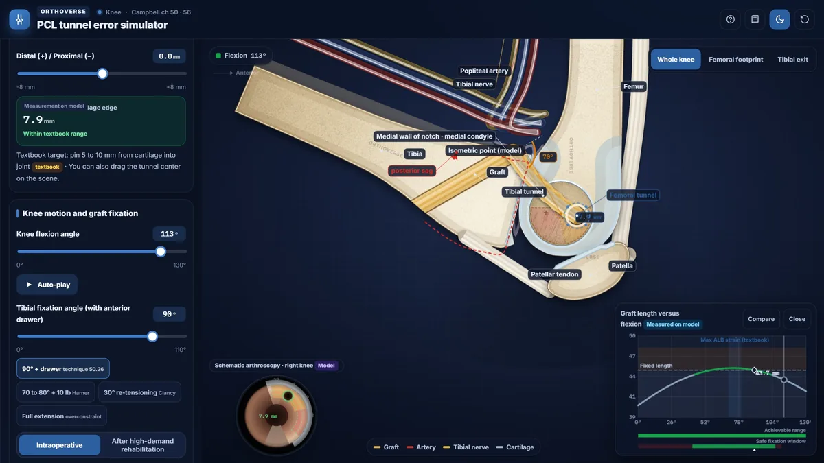
- Load the anatomic ALB center, the isometric point and the two classic errors: anterior-distal and posterior-proximal.
- Compare fixation angles: 90° with an anterior drawer, 70° to 80° with 10 lb, 30° re-tensioning, and full extension.
- Switch between transtibial and tibial inlay and see the killer turn at the tibial exit; change the tunnel angle.
- See the popliteal artery and tibial nerve on the same view, and work through board exam traps.
Key concepts
The PCL has a large anterolateral bundle and a smaller posteromedial bundle; most fibers lengthen as the knee flexes, and the femoral tunnel position matters more than the tibial one.
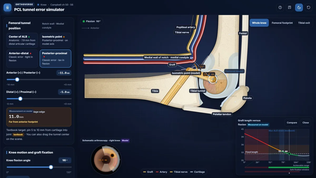
A tunnel that is too anterior and distal tightens the graft in flexion and limits it; a posterior and proximal tunnel leaves the graft lax in flexion, with posterior sag.
The transtibial graft must turn sharply at the back of the tibia (the killer turn); a tibial inlay or a careful tunnel angle reduces it.
Teaching notes
The posterior cruciate ligament (PCL) consists of a large anterolateral bundle (ALB) and a smaller posteromedial bundle (PMB); the ALB is the primary restraint to posterior tibial translation and is tightest near 70-80 degrees of flexion. Accurate femoral tunnel placement on the lateral wall of the medial condyle is the single most consequential technical decision in PCL reconstruction, because even small errors in the proximal-distal direction alter graft length patterns far more than equivalent errors in the anterior-posterior direction.
Femoral Footprint and Tunnel Target
The PCL femoral footprint lies on the lateral surface of the medial condyle and is approximately semicircular. The center of the ALB sits 7.9 mm from the distal articular cartilage. Arthroscopically in a right knee, the isometric point is 8 mm proximal to the articular cartilage at the 1 o'clock position. During drilling, the guide pin should enter 5 to 10 mm from the intra-articular cartilage, and 1 to 2 mm of bone must remain at the articular margin after reaming.
A key asymmetry governs tunnel placement errors: a proximal-distal shift alters the graft length pattern approximately three times as much as an equivalent anterior-posterior shift. Campbell therefore states that if any error must occur, it should be in the anterior-posterior direction rather than the proximal-distal direction.
Classic Femoral Tunnel Errors
An anterior-distal femoral tunnel increases the distance between the two tunnel centers as the knee flexes, so the graft tightens progressively in flexion. The clinical result is loss of 10 to 20 degrees of flexion; if the surgeon attempts to force motion, the graft is overstretched.
A posterior-proximal femoral tunnel has the opposite effect: the inter-tunnel distance decreases with flexion, leaving the graft lax throughout the flexion arc. The tibia sags posteriorly, and posterior sag and drawer persist. On stress view, residual posterior translation greater than 4 mm defines residual posterior laxity.
Tibial Tunnel and the Killer Turn
In the transtibial technique, the graft exits the posterior tibial aperture and turns sharply toward the femoral tunnel, forming a near-right-angle bend known as the killer turn. This bend abrades and thins the graft and complicates tensioning. The tibial guide should be angled approximately 60 degrees relative to the tibial articular surface; a more perpendicular angle creates an excessively acute bend at the posterior tibia, and a tunnel started too distally may ream out the posterior tibial shelf.
Two strategies reduce the tibial-side bend: the tibial inlay technique places a bone block in the posterior trough, eliminating the exit bend and allowing a larger graft; alternatively, an anterolateral tibial tunnel starting point reduces the wrap angle. Neither approach has been proven superior in clinical outcome studies.
Graft Fixation Angle
The fixation angle determines the reference length against which all knee positions are compared. Fixation in full extension without load creates an overconstrained knee: every degree of flexion demands additional graft length beyond the fixed length, generating high graft forces and reducing total translation. Fixation at 90 degrees of flexion with an anterior drawer produces kinematics that most closely approximate the normal knee.
Two established techniques reflect this principle: in the standard technique the tibia is held at 90 degrees with an anterior drawer; in the Harner technique an Achilles allograft is fixed at 70 to 80 degrees with 10 pounds of tension, because the ALB is under maximal tension in that range.
Neurovascular Safety During Tibial Drilling
The popliteal artery and tibial nerve are at risk when the tibial tunnel is drilled. The anterior wall of the popliteal artery lies only 7 to 10 mm posterior to the PCL; this distance increases with knee flexion. Drilling at 90 degrees of flexion moves the neurovascular bundle away from the tibia. A curette or protective guide should be placed posterior to the tibia before pin advancement, and fluoroscopy should confirm pin position before reaming.
Postoperative Considerations
After PCL reconstruction, the knee is braced with the hinge locked in full extension. An extension deficit or flexion contracture appearing weeks to months postoperatively is usually caused by prolonged immobilization in flexion, not by tunnel malposition; tunnel errors principally affect either flexion range or posterior stability, not extension. A late complication of femoral-side drilling is osteonecrosis of the medial condyle, arising from subchondral vascular injury during dissection or reaming.
Try this in the simulator
- Drag the tunnel center to the ALB center position and open the Graft length vs. flexion chart to see the graft tighten near 70-80 degrees, matching native ALB behavior.
- Shift the tunnel anterior and distal, then use Auto-play to flex the knee and observe when Flexion blocked appears and the range of motion reading drops.
- Move the tunnel posterior and proximal and switch the Outcome to After high-demand rehabilitation to see how graft creep produces residual posterior laxity on the simulated stress view.
- On the Tibial side panel, switch between Transtibial and Tibial inlay while watching the Bend angle and Abrasion model indicators change.
- Set Tibial fixation angle to Full extension and compare the graft length curve with the fixed length dashed line, then change to 90 degrees with drawer and note how the overconstrained pattern resolves.
Pitfalls
- Anterior-distal femoral tunnel is the classic board trap for loss of flexion after PCL reconstruction; the inter-tunnel distance increases with flexion and the graft becomes overtight.
- Posterior-proximal femoral tunnel leaves the graft lax throughout the flexion arc; the posterior drawer and sag are not controlled, and residual laxity exceeds 4 mm on stress view.
- Proximal-distal tunnel error is roughly three times more damaging to graft length behavior than an equivalent anterior-posterior error; always err in the AP direction.
- Fixation in full extension without load overconstrains the knee and generates high graft forces; fix at 90 degrees with anterior drawer or 70-80 degrees with 10 pounds.
- Extension deficit after PCL reconstruction is usually from prolonged immobilization in flexion, not tunnel malposition; do not assume a tunnel error when flexion contracture appears.
One-page high-yield card
The key facts, numbers and pitfalls of this simulator on one printable page. Free for personal study.
Further reading
- Campbell's Operative Orthopaedics, 15th edition (2026): Knee Injuries, ch 50 (PCL anatomy, reconstruction techniques, fixation and complications).
- Campbell's Operative Orthopaedics, 15th edition (2026): ch 56 (arthroscopic PCL technique).

Designed for Orthoverse by Dr. M. M. Bakhshaei. Built to make orthopedic concepts visible, measurable and easy to explore.
Have an idea for the next simulator?
Tell us which concept you would like to explore. Message our bot and we will read every idea.
For medical education only. Orthoverse simulators are teaching models for health professionals and students. They are simplified on purpose, are not medical devices and are not a substitute for clinical judgement, patient-specific imaging or the original literature.
© 2026 Orthoverse. All rights reserved. The simulators, their code, drawings and models are original work. Free to use for learning on orthoverse.org; copying, embedding, scraping or redistribution is not permitted. Textbook references are cited for further reading only.