
Plate and Screw Strain Lab
Perren's interfragmentary strain theory, live: choose the fracture pattern, the plating method, the screws and the material, change the gap and the load, and watch strain cross the tolerance of bone, cartilage and granulation tissue.
What you can do
- Pick the fracture pattern: transverse, oblique, wedge or comminuted.
- Choose compression, lag screw plus neutralization, or a bridge plate, with conventional or locking screws in steel or titanium.
- Change the gap, the working length (screw positions), the plate offset from bone and the load.
- Read interfragmentary strain on a gauge with the tolerance bands of bone, cartilage, transition and granulation tissue.
- Load seven classic cases, from compression of a simple fracture to locking off the bone.
- See strain under the plate and opposite the plate, and the expected type of healing.
Key concepts
Absolute stability (anatomic reduction, compression, rigid fixation) gives primary bone healing; relative stability gives healing with callus.
Strain is gap motion divided by gap width: the same motion is tolerable across a wide comminuted zone but too much for a narrow simple fracture gap.
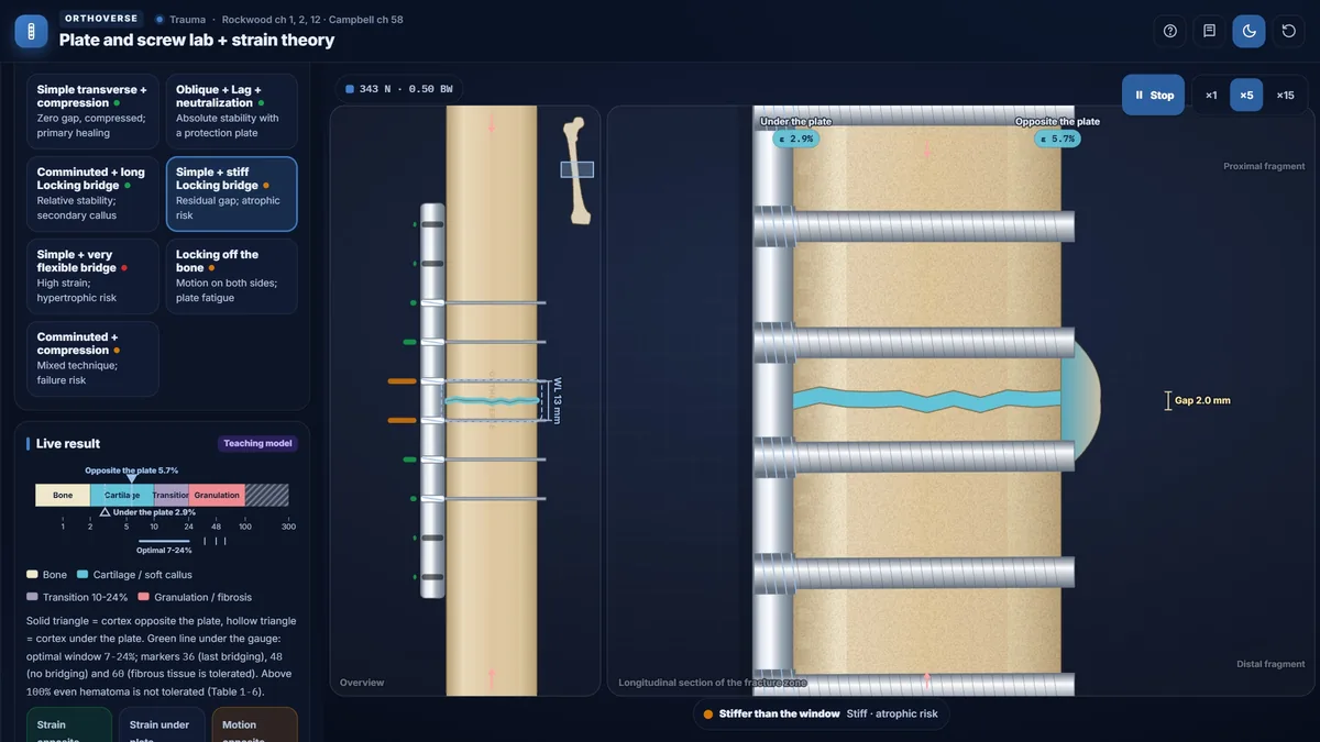
A stiff bridge over a simple fracture leaves a small gap with too little strain, a classic route to atrophic nonunion; a plate held far from the bone moves on both sides of the fracture and fatigues.
Teaching notes
Interfragmentary strain governs whether a fracture heals by direct bone remodeling or by callus formation. Understanding the relationship between gap size, fragment motion, and tissue tolerance allows the surgeon to choose a fixation strategy that matches the mechanics to the biology.
Strain Theory
Perren's strain is interfragmentary motion divided by gap width. The same absolute motion produces high strain in a narrow simple-fracture gap and proportionally lower strain spread across a wider comminuted zone. Tissue tolerates strain only up to a characteristic limit: hard callus (bone) up to 2%, soft callus up to approximately 10 to 12.8%, and hematoma up to about 100%. The classic callus-forming range is 2 to 10%; the optimal window from sheep studies is 7 to 24%.
- Strain = interfragmentary motion ÷ gap
- Hard callus (bone) limit: 2%; soft callus: 10–12.8%; hematoma: ~100%
- Classic callus range: 2–10%; optimal window: 7–24%; bridging absent above 48%
Primary and Secondary Healing
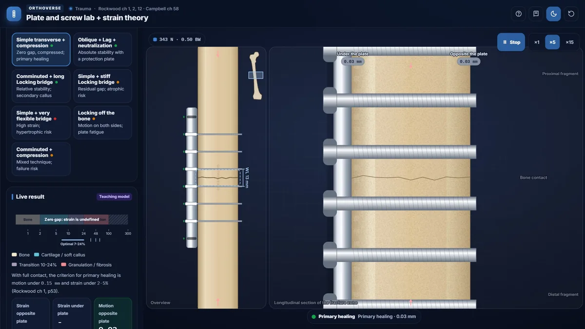
Primary (direct) healing requires anatomic reduction, compression, and rigid fixation with motion under 0.15 mm and no residual gap. Osteons cross the interface by remodeling and no periosteal callus appears on radiographs. Secondary healing follows relative stability: a residual gap and cyclic motion stimulate periosteal callus, which forms predominantly on the cortex opposite the plate where strain is higher. Combining a non-anatomic reduction with absolute-stability fixation satisfies neither criterion and frequently leads to failure.
- Primary healing: zero gap + compression + motion < 0.15 mm
- Secondary healing: relative stability, residual gap, callus on the side opposite the plate
- Non-anatomic reduction + rigid fixation = neither primary nor secondary healing
Plating Methods and Working Length
Three plating methods serve three biomechanical goals. Compression plating achieves absolute stability for simple transverse or short oblique fractures with anatomic reduction; slight plate overcontouring keeps the far cortex in contact. Lag screw plus neutralization achieves absolute stability for oblique fractures by compressing the oblique surface and protecting against rotational and bending loads. Bridge plating spans a comminuted zone with relative stability, relying on callus for healing, using a long plate with few screws and a deliberately long working length.
Working length is the distance between the two innermost screws on either side of the fracture. Construct axial stiffness falls approximately as the square of the ratio of the original to the new working length. A longer working length is intentional in bridge plating; an inadvertently long working length in a simple fracture creates strain the narrow gap cannot tolerate.
Screw Configuration and Plate Fatigue
The screws nearest and farthest from the fracture contribute most to stiffness; intermediate screws add progressively less. At least six to eight cortices per side are generally recommended; a locking plate requires a minimum of two screws in each fragment. Locking screws thread into the plate hole at a fixed angle and fail in parallel rather than sequentially. Plate strain is highest at the two holes immediately beside the fracture and falls with distance; an empty hole directly over the fracture substantially raises plate fatigue failure risk.
- Minimum 6–8 cortices per fragment; at least 2 locking screws per fragment
- Locking screws act in parallel and pull out simultaneously
- Highest plate strain: two holes beside the fracture; empty hole over fracture raises fatigue risk
Nonunion Patterns
Atrophic nonunion follows a stiff construct with a residual gap: strain is nearly zero, providing no callus stimulus, and the gap is too wide for direct bone crossing. Hypertrophic nonunion follows excessive motion: abundant callus forms but cannot bridge the gap because strain exceeds the tolerance of mature repair tissue. Moving a locking plate away from the bone increases motion on both sides and reduces the fatigue load the construct can sustain.
- Atrophic nonunion: stiff fixation + residual gap → near-zero strain, no callus
- Hypertrophic nonunion: excessive motion → callus without bridging
- Titanium is approximately half as stiff as steel; lower stiffness shifts the construct toward the callus-forming range
Try this in the simulator
- Load the 'Simple transverse + compression' preset: confirm the gap is zero and the teaching model shows primary healing.
- Switch to 'Comminuted + long Locking bridge' and observe how comminution distributes motion so strain falls into the callus range.
- Select 'Simple + stiff Locking bridge' and note how a residual gap with near-zero strain places the construct in the atrophic nonunion zone.
- Use the Plate holes screw controls to remove intermediate screws; watch Axial stiffness and the Interfragmentary strain gauge respond.
- Load 'Locking off the bone', increase Plate offset from bone, and compare Strain under plate with Strain opposite plate.
Pitfalls
- Applying rigid fixation to a non-anatomic reduction: bone cannot be compressed and callus cannot form, a classic route to failure.
- Leaving an empty hole over the fracture with a bridge plate: plate strain peaks at the adjacent holes and fatigue failure risk rises substantially.
- Using a locking plate for compression without first applying compression with a conventional screw or device; locking itself does not compress.
- Short working length on a bridge plate: high stiffness reduces the callus stimulus and risks atrophic nonunion.
- Confusing nonunion types: hypertrophic (excess motion, no bridging) requires stiffer fixation; atrophic (gap + no callus) requires biological intervention.
One-page high-yield card
The key facts, numbers and pitfalls of this simulator on one printable page. Free for personal study.
Further reading
- Rockwood and Green's Fractures in Adults, 10th edition: ch 1, 2 and 12 (biomechanics, bone healing and principles of fracture fixation).
- Campbell's Operative Orthopaedics, 15th edition (2026): ch 58 (general principles of fracture treatment).

Designed for Orthoverse by Dr. M. M. Bakhshaei. Built to make orthopedic concepts visible, measurable and easy to explore.
Have an idea for the next simulator?
Tell us which concept you would like to explore. Message our bot and we will read every idea.
For medical education only. Orthoverse simulators are teaching models for health professionals and students. They are simplified on purpose, are not medical devices and are not a substitute for clinical judgement, patient-specific imaging or the original literature.
© 2026 Orthoverse. All rights reserved. The simulators, their code, drawings and models are original work. Free to use for learning on orthoverse.org; copying, embedding, scraping or redistribution is not permitted. Textbook references are cited for further reading only.